HomePopularColourful Scans Show the Millions of Bacteria Lurking on Human Hair and...
Colourful Scans Show the Millions of Bacteria Lurking on Human Hair and Skin
6585
Coloured scanning electron micrograph (SEM) of Photocomposite, Staphylococcus aureus on the surface of the small intestine villi. Gram-positive, MRSA, coccus prokaryote (dividing); causes food poisoning, toxic shock syndrome and skin and wound infections such as scalded skin syndrome, scarlet fever, erysipelas and impetigo. Dennis Kunkel Microscopy/ SPL / mediadrumworld.com
Bacteria in Humans
By Rebecca Drew
STOMACH-CHURNING images have revealed the gruesome bacteria lurking on human hair and skin and hiding inside the body.
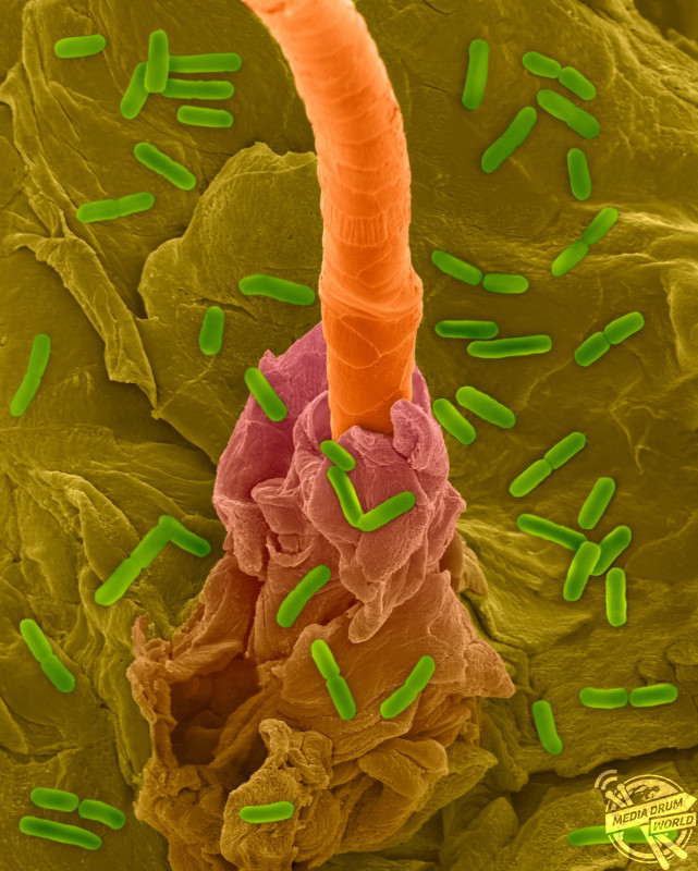
Coloured scanning electron micrograph (SEM) of Photocomposite, E. coli on the surface of human skin and hair follicle. Dennis Kunkel Microscopy/ SPL / mediadrumworld.com
The colourful scans show E. coli dotted on the skin’s surface and around human hair follicles. Other fascinating shots show the bacteria of a human lung infection and bright green shots show the Streptococcus salivarius that causes plaque on teeth.
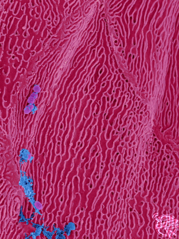
Coloured scanning electron micrograph (SEM) of Bacteria on an epithelial cells from the human tongue filiform papillae. Dennis Kunkel Microscopy/ SPL / mediadrumworld.comColoured scanning electron micrograph (SEM) of Photocomposite of bacteria on human skin. Dennis Kunkel Microscopy/ SPL / mediadrumworld.com
One eye-catching picture shows food poisoning causing bacteria in the small intestine.
The interesting scans were taken by Hawaii based award-winning microscopist, Dennis Kunkel.
Coloured scanning electron micrograph (SEM) of Streptococcus salivarius, Gram-positive, coccoid, facultatively anaerobic prokaryote (bacterium). Dennis Kunkel Microscopy/ SPL / mediadrumworld.com
“I worked with some researchers in the nineties on human skin and intestinal bacteria and continued to obtain bacteria from these areas to study and photograph for my image collection,” said Dennis.
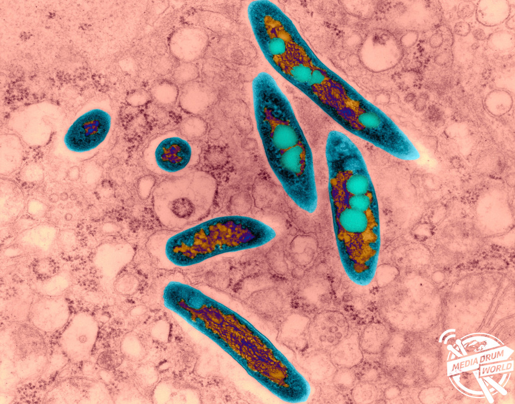
Coloured transmission electron micrograph (TEM) of Mycobacterium avium complex (MAC) infection (human lung). Dennis Kunkel Microscopy/ SPL / mediadrumworld.com
“I have worked with many microbiologists and clients who have supplied human bacteria.
“Using electron microscopy methods and imaging reveals many things about bacterial morphology and human bacterial interactions.”
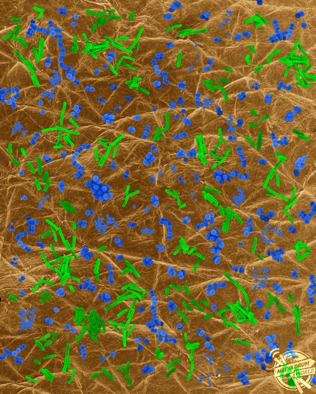
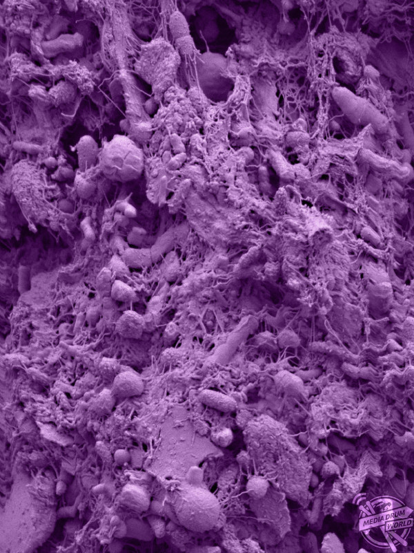
Coloured scanning electron micrograph (SEM) of Human intestinal bacteria (small intestine). This image shows the human gut epithelia with symbiotic microbes that interact to maintain health. Dennis Kunkel Microscopy/ SPL / mediadrumworld.com
“The microbiological community diversity interests me the most in the human intestine and skin.”
According to researchers at the Weizmann Institute of Science in Rehovot, Israel, there is a ratio of 1.3 bacteria to each human cell.
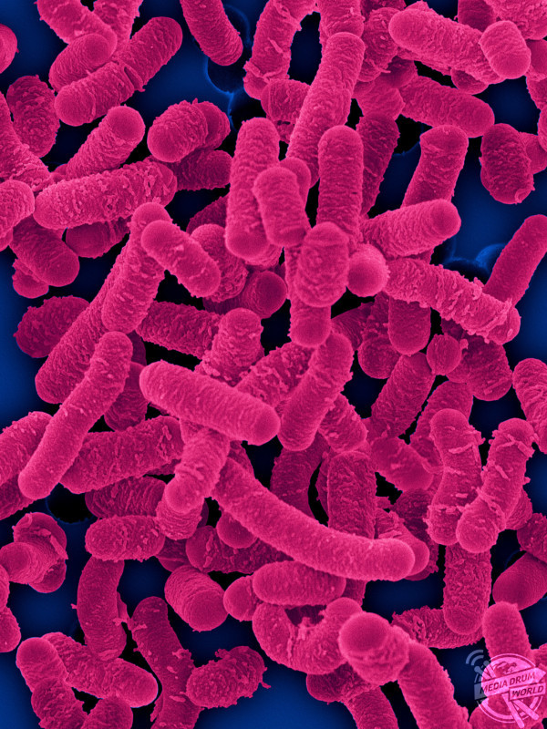
Coloured scanning electron micrograph (SEM) of Lactobacillus salivarius is homofermentative, probiotic bacterium that lives in the human gastrointestinal tract. Dennis Kunkel Microscopy/ SPL / mediadrumworld.com